What a cell line is,and why it matters.
Almost everything we offer depends on one object: a vial of living cells. This essay explains what that object is, how it is made, and what it can and cannot do.
Reem Bio Laboratory ·
i.
A cell carries everything
Every animal is built from cells, and almost every one of those cells carries a complete copy of the animal's genome – the full sequence of DNA inherited from its mother and father. A skin cell from a dog's ear holds the same genetic instructions as a cell from its heart or its brain. What differs between them is which instructions are being used. This is the quiet fact on which cell banking rests: to keep a genome, you do not need the whole animal, or even a large part of it. You need a few living cells.
The difficulty is the word living. DNA itself is a robust molecule – it can be extracted from hair, blood or saliva and stored dry for years. But extracted DNA is a record, not a source. It can be read; it cannot be grown, and it cannot be used to make new cells. A living cell can. It can divide, it can be coaxed into producing more of itself, and – in the case of cloning – its nucleus can direct the development of a whole new animal. The aim of preservation is therefore not to store DNA, but to store cells that are still alive and able to divide.
ii.
From biopsy to line
A cell line begins with a biopsy: a piece of skin a few millimetres across, taken by a veterinarian under local anaesthetic. In the laboratory, the tissue is washed, cut into fragments of about a millimetre, and laid in a culture dish with a nutrient medium warmed to body temperature. These fragments are called explants. For the first day or two nothing seems to happen. Then, at the edges of each fragment, cells begin to creep out onto the plastic.
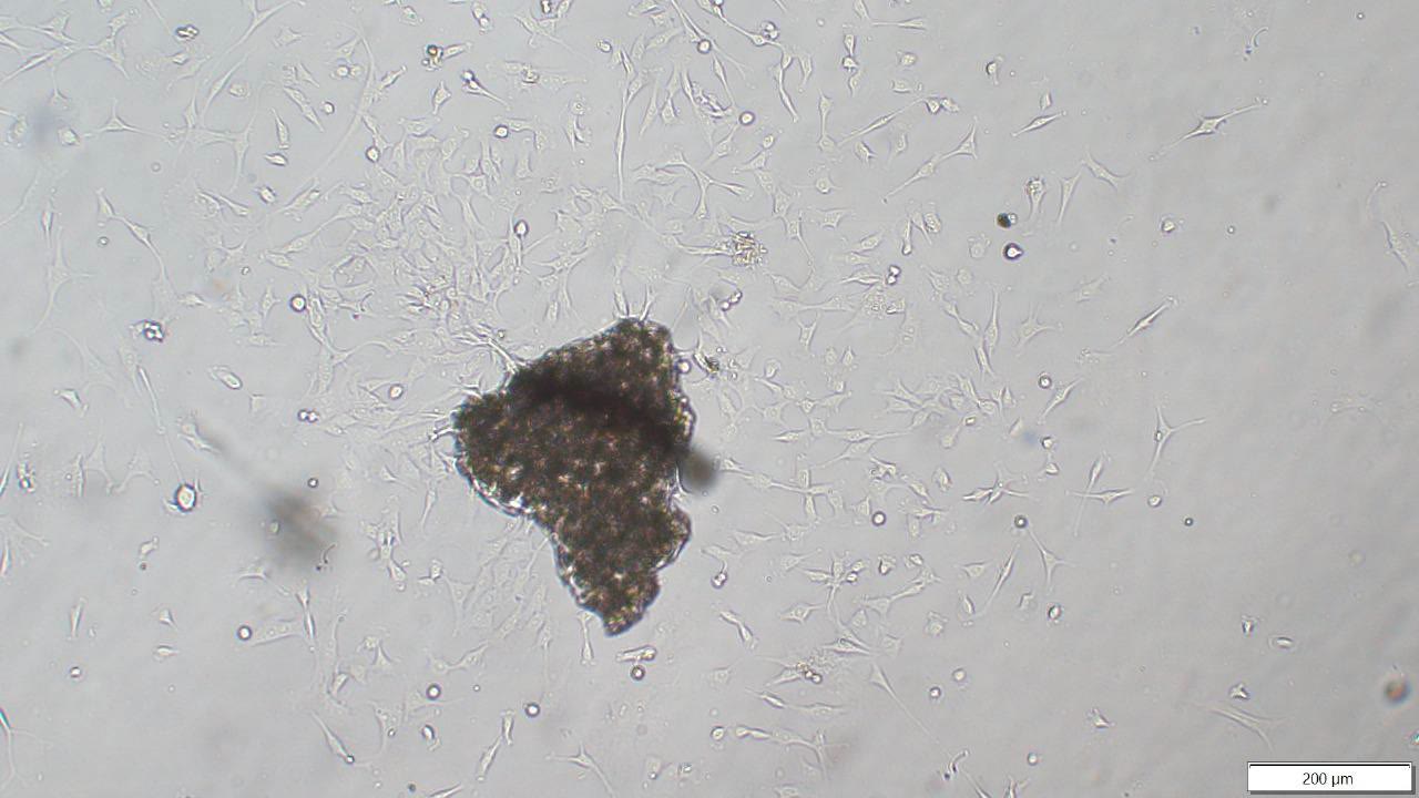
These cells are fibroblasts, the workhorses of connective tissue. They are the cells that heal a wound, and they are good at the laboratory equivalent: leaving the tissue, attaching to a surface, and dividing. Over the following two weeks they multiply until they cover the dish. At that point, called confluence, they are gently detached and spread across larger flasks. Each round of this is called a passage. After two or three passages, there are enough cells – many millions – to freeze a full deposit.
We freeze cells early, at low passage, for a reason that has been understood since the 1960s. Ordinary cells taken from a body can only divide a limited number of times before they slow and stop, a phenomenon named after the biologist who described it. Freezing a line young keeps as much of that capacity in reserve as possible.²
iii.
Freezing without harm
Cells are mostly water, and water that freezes forms crystals that can tear a cell apart. The art of cryopreservation is to cool cells in a way that avoids this. Before freezing, the cells are suspended in a medium containing a cryoprotectant, a compound that slips into the cell and limits the formation of ice. They are then cooled slowly – about one degree a minute – which gives water time to leave the cell before it can freeze inside it. The physics of this was worked out in detail in the second half of the twentieth century and remains the basis of cell banking today.¹
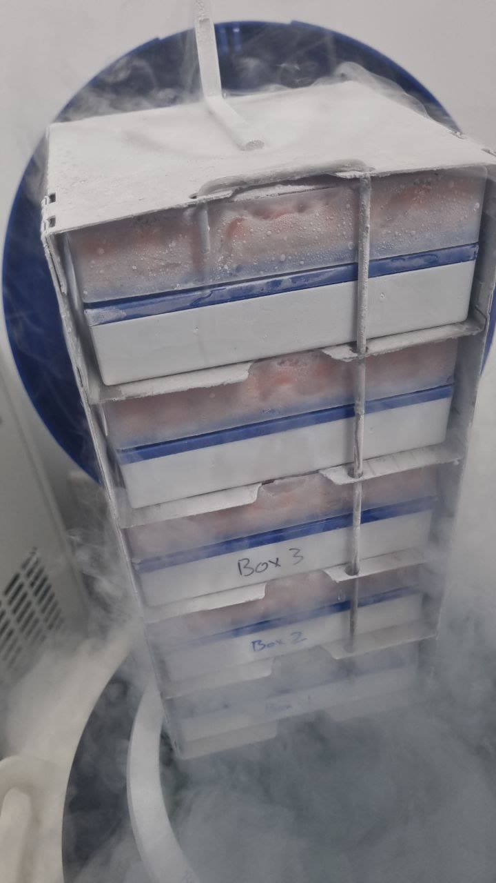
Once the cells reach around −80 °C, they are moved into the vapour above liquid nitrogen, where the temperature sits well below −150 °C. At these temperatures the chemistry of the cell effectively stops. There is no metabolism, no ageing, no decay. A vial stored correctly today should, as far as anyone has been able to observe, be the same vial in thirty years. We are careful not to claim more than has been observed; but there is no known mechanism by which cells held at this temperature deteriorate.
iv.
Testing before trusting
A cell line is only as useful as it is clean and alive. Before any line is banked, we test it for mycoplasma, a group of very small bacteria that can infect cultures without any visible sign, and for bacterial and fungal contamination. We record a DNA identity profile so that the line can always be tied to the animal it came from. And we thaw one vial from every deposit to count how many cells survive – the viability test. Only when all of these pass is a certificate issued.
Then the deposit is divided. Half of the vials go to the primary vault, usually in Abu Dhabi. Half go to a mirror in another city, usually Bahrain. Each site holds enough to rebuild the line alone. This is not a precaution against something we expect; it is a precaution against everything we do not.
v.
Why it matters
A cell line is valuable because it keeps choices open. From it, a laboratory can extract DNA for genetic testing at any time, as often as needed, without disturbing the animal. It can grow stem cells or other cells for regenerative therapies prescribed by a veterinarian. And it can provide the nucleus for cloning, producing a genetic twin years or decades later.
But the most important thing a cell line does is let an owner separate two decisions that are often confused. The first – whether to keep the possibility – has to be made while the animal is alive, or very shortly after. The second – what, if anything, to do with it – can be made at leisure, with better information, when grief or urgency have passed. Many of the lines in our vaults will never be used for anything, and that is a good outcome. They were kept so that the decision could be made well.
- How big is the biopsy?
- A piece of skin a few millimetres across, taken under local anaesthetic by a veterinarian. Most animals are home or back in the stable within the hour.
- What if the cells do not grow?
- Roughly one sample in twenty needs a second biopsy. We tell you as soon as we see it, and the repeat is not charged.
- Is a cell line the same as a DNA sample?
- No. Extracted DNA can be read but not grown. A cell line is alive, and can supply DNA, cells for therapy or a nucleus for cloning.
- Do I have to use the line?
- No. Preservation is complete in itself. Many lines are never used, and that is a good outcome.
Ask us about your animal's line.
Book a consultation